UNOSSIFIED NASAL BONE / ABSENT NASAL BONE IN SCAN – WHAT DOES IT MEAN?
Dr. Zohra Ahmad
Fetal Medicine Specialist
Institute of Human Reproduction
The nasal bone is a paired bone forming the anterior wall of the nasal cavity and is largely responsible for the shape of the nose. The nasal bones are variable in size and form in different individuals. The first trimester scan is an important tool of risk assessment for fetal aneuploidies. The presence or absence of fetal nasal bone is an important secondary marker for risk allocation.
1. What is Unossified / Absent Nasal Bone?
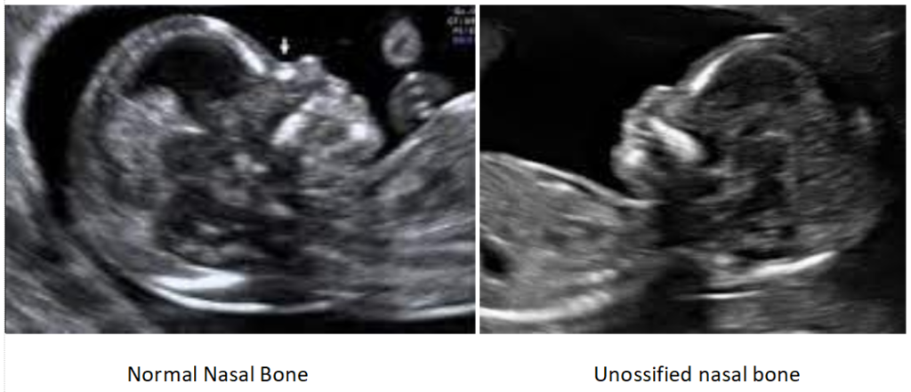
It refers to a finding on ultrasound that nasal bone is not seen or appears small. It is due to delayed ossification of the nasal bone. In the report, it may be mentioned as either unossified, absent, or hypoplastic nasal bone. Unossified and absent nasal bone means that nasal bone is not seen while Hypoplastic nasal bone means nasal bone is seen but very small.
2. Causes of Absent Nasal Bone
It can be due to hypoplasia or delayed ossification of the nasal bone. It can be due to underlying chromosomal abnormality or in 1-2% of normal babies as well.
3. How Common Is Absent Nasal Bone?
It is found in 1-2% of normal babies. Studies have shown that in about 85-90% cases of the isolated absent nasal bone, babies will be normal. However, in about 10-12% of cases, babies may have chromosomal problems including down’s syndrome .
4. When can Unossified Nasal bone be detected?
Unossified nasal bone in the first trimester
It is generally detected in the first trimester that is 11-14 weeks in a special ultrasound scan called First trimester anomaly scan/ First trimester scan or NT/NB scan. It must be done by an expert radiologist or fetal medicine specialist with special training, using a high end ultrasound machine with baby in appropriate position.
Unossified nasal bone in second trimester/20 weeks scan or third trimester
Sometimes if the first trimester scan has not been done, unossified nasal bone can also be first detected later in the second or third trimester. The significance of absent nasal bone in different trimesters is the same.
5. What is the Significance of detecting Unossified Nasal Bone?
It can be a soft marker of Down’s syndrome and other chromosomal abnormalities. Soft marker means any ultrasound sign which increases the likelihood of your baby having Down’s syndrome. More than half of babies with chromosomal abnormalities may have absent nasal bone. Normally every human being has 46 chromosomes or 23 pairs of chromosomes.
The most common chromosomal problem encountered is Down’s syndrome which is an aneuploidy of chromosome 21 where instead of two chromosomes, three are present. To know more about common aneuploidies, click the linked article. Affected children are mentally challenged and may have other health issues like heart defects and duodenal atresia. They also require special schools. However, it is important to note that 1-2% of normal babies may also have an unossified nasal bone. Hence there is no need to be alarmed when your pregnancy scan reports an unossified nasal bone.
6. What if I had an unossified nasal bone in the first-trimester scan and it ossifies in the second trimester or anomaly scan
This is a common situation. Once the unossified or hypoplastic nasal bone has been identified the risk for down’s syndrome /other aneuploidies remains the same even if it ossifies at a later stage.
7. What Next?
If unossified nasal bone is detected in the first trimester, your radiologist or fetal medicine specialist will provide you one of the following options to assess your risk of a chromosomally abnormal baby.
Combined first-trimester screening/ Quadruple screening:
These are blood tests done in first and second trimester respectively. Sensitivity of first trimester combined screening for detecting aneuploidies is 90-95 % while of quadruple test is 60-70%. These test results can be either low risk or high risk. If low risk, then it is reassuring and we can follow up the pregnancy. If high risk, a definitive test like Chorionic villus sampling or amniocentesis for karyotyping the baby is advisable.

Non-invasive prenatal testing (NIPT):
This is a blood test based on cell free fetal DNA which is basically DNA of the baby that circulates in maternal blood. It has a high sensitivity of 99%. Result can be low risk, high risk or no call. If low risk, pregnancy can be followed up. If high risk or no call—we have to go for direct testing.

Direct testing:
Can be either Chorionic villus sampling or Amniocentesis which is done to get the karyotype or the chromosomal make-up of the baby. In Chorionic villus sampling, tissue is taken from the placenta, while in Amniocentesis, around 20-30ml fluid is taken from around the baby. These tests are generally safe in expert hands however may carry a procedure related risk of miscarriage of around 0.3 %.

8. What is the further course in Pregnancy?
If the risk for having a chromosomal abnormality based on the above tests is found to be low or if karyotype shows normal result then we will routinely follow up the pregnancy at 20 weeks for anomaly scan and growth scans thereafter.
If Karyotype is abnormal, say the baby has Down’s syndrome or any other chromosomal abnormality, parents can be given an informed choice of pregnancy termination.
9. Does Absent / Unossified Nasal bone affect the baby in any way after birth? Will my baby’s face have any cosmetic problem?
No. Unossified nasal bone is only an aneuploidy marker or a soft sign for Down’s syndrome. In the absence of Down’s syndrome, it does not affect the baby in any way.
10. Is any treatment required for unossified nasal bone
No. treatment is not required as it ossifies after birth.
20 week anomaly scan is done but not seen nasal bone everything part is babies normal 13 week nt scan also done doctor suggest dual marker that also done report is low risk but I m worried about it doctor suggest NIPT
PLEASE GIVE ME ANY SOLUTION FOR BABY NASAL BONE
Mere bachhe ke naak ki haddiya abhi nahi bani he to me kya karu
Can you tell us more about this? I’d love to find out some additional information.
Here is my blog … y2mat e
“I had PCOD. My doctor put me on medication for three months, and then I got pregnant. My last date was 30/9/2025. From the beginning I took folic acid and got all scans done. My first scan where they check the heartbeat, NT/NB scan, double marker test, and anomaly scan — till now everything came normal. But yesterday I got my 7th month Doppler scan done, and in that the baby’s right kidney is not visible and there is a two-vessel umbilical cord. Then the doctor told me yesterday itself to get a scan done at another place for a cross-check. In that report it came that nasal bone is unossified, right kidney not seen, single umbilical artery, and the baby’s position is breech. Ma’am, I don’t understand why all this didn’t show up in earlier scans and why it has suddenly happened now. My anomaly scan was done in Allahabad and Doppler in Indore. Ma’am, could there be danger to the baby? Rest of the things — blood flow, amniotic fluid, heartbeat, movement — are normal in the baby.” In my nt scan nasal bone was shown present and artery was also normal same with anamoly scan then why in doppler scan it is showing that my baby is missing these things and what possible defects my baby may have.
Dear Pooja, we can truly feel the anxiety you are going through.
To answer your question on ‘why now’: Anomaly scans are usually done between 18–22 weeks. As the baby grows into the third trimester, certain structures (like the kidneys or the complexity of the heart and blood vessels) become more prominent. Sometimes, a finding like a Single Umbilical Artery (SUA) or a non-visualized kidney can be missed earlier due to the baby’s position or may only become clear as the baby’s size increases.
Please meet the following specialists as soon as possible.
1. Fetal Medicine Specialist: Since you have had two different reports, we highly recommend seeing a certified Fetal Medicine specialist
2. Genetic Counseling: A specialist can help you decide if further tests (like a late-term amniocentesis) are necessary based on the combination of these markers.
3. Consult a Pediatric Surgeon: If the single kidney is confirmed, meeting a specialist now will help you understand the management usually is after birth.
Please stay strong.
Best wishes.
Dr. Deepak Goenka
Director, IHR Guwahati